
Figure 3.
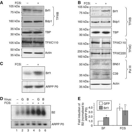
Brf1 levels limit pol III transcription in unstimulated cardiomyocytes, but increase specifically after hypertrophic stimulation. (A) Whole-cell lysates of rat1A fibroblasts that had been serum-starved for 24 h then either maintained in serum-free media or exposed to 10% FCS for 16 h were resolved by SDS–PAGE, and immunoblotted using antibodies against the proteins indicated. (B) The same procedure as outlined in (A) was used to analyse expression of the indicated proteins in cardiomyocytes. (C) Cultured cardiomyocytes were treated as in (A), then total RNA was extracted and used to generate cDNAs, which were amplified by PCR using primers specific for Brf1 or ARPP P0. (D) Cultured cardiomyocytes were infected with adenoviruses expressing HA-Brf1 (B) or GFP (G). Cells were serum-starved or stimulated with 10% FCS for 16 h before extracting whole-cell RNA. RNA was analysed by Northern blotting using B2- and ARPP P0-specific probes. (E) Northern blots were quantified by densitometry and B2 levels normalised to ARPP P0. The fold increases in normalised B2 expression relative to serum-starved, GFP control infected cells are shown. (n=8; *significantly different from SF GFP, P<0.05; **not significantly different from FCS GFP.)