
Figure 7.
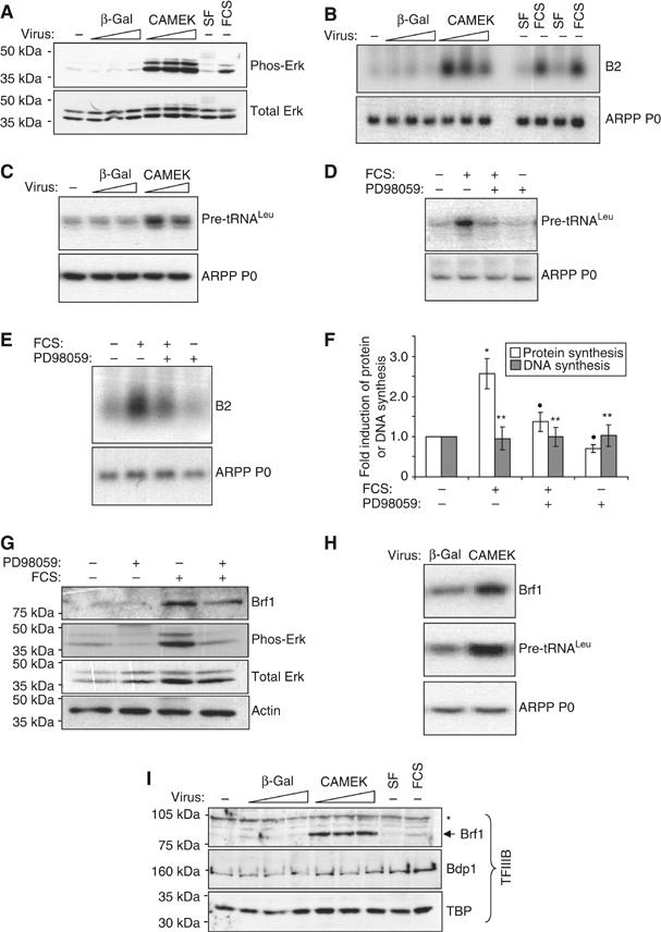
Erk induces pol III transcription and Brf1 expression in cardiomyocytes. (A) Cultured cardiomyocytes were infected (at a multiplicity of infection (m.o.i.) of 20, 50 or 80) with adenovirus expressing CAMEK or β-gal (negative control), as indicated. Cells were serum-starved for 48 h before harvesting. As a positive control for Erk activation, uninfected cells were incubated without (SF) or with 10% FCS for 16 h. Whole-cell protein was analysed by Western blotting using the antibodies indicated. (B) Total RNA from cells treated as in (A) was analysed by Northern blotting using the indicated probes. (C) Infections were carried out as in (A), using an m.o.i of 50 or 80, then total RNA analysed by RT–PCR using pre-tRNALeu- and ARPP P0-specific primers. (D–G) Cells were cultured in the absence of serum or stimulated with 10% FCS for 16 h. PD98059 or vehicle were also included for 16 h where indicated. Whole-cell RNA was analysed by (D) RT–PCR or (E) Northern blotting. (F) Protein and DNA synthesis rates were determined by measuring the incorporation of [35S]methionine/cysteine or [3H]thymidine, respectively. The fold increases represent the mean of four separate experiments, each with three replicates per condition (*significantly higher than SF+vehicle, P<0.05; •significantly lower than FCS+vehicle, P<0.05; **not significantly different from SF+vehicle). (G) Protein lysates were analysed by Western blotting using antibodies against the proteins indicated. (H) Total RNA was extracted from cardiomyocytes infected (at an m.o.i of 50) with adenovirus expressing CAMEK or β-gal (negative control), as indicated. Cells were serum-starved for 48 h before harvesting. RT–PCR was performed using the primers specified. (I) Whole-cell lysates, treated as in (A), were analysed by Western blotting using the indicated antibodies (*nonspecific band).