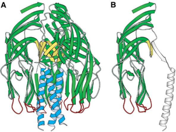
Figure 2.

VP5CT and the VP5* antigen domain. (A) Ribbon diagram of the VP5CT trimer, colored to match Figure 1. VP5CT does not include the foot region. (B) Ribbon diagram of a single VP5CT subunit. The part that forms the VP5* antigen domain is green, yellow, and red; the remainder is drawn in outline.
