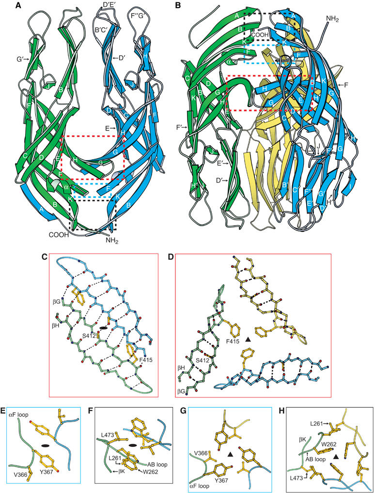
Figure 3.

Crystal structures of the VP5* antigen domain dimer and trimer. (A) Ribbon diagram of the dimer. The orientation matches the model of the rigid spikes on the virion in Figure 1A. The red, blue, and black boxes show the regions detailed in panels (C), (E), and (F), respectively. The termini of the green subunit are indicated. Secondary structural elements, including the three hydrophobic loops of the blue subunit are labeled. (B) Ribbon diagram of the trimer. The orientation matches the model of the rearranged trimer in Figure 1B. The red, blue, and black boxes show the regions detailed in panels (D), (G), and (H), respectively. The termini of the blue subunit are indicated. (C, E, F) Atomic details of the key intersubunit contacts of the dimer. Black ovals indicate the approximate two-fold axis. In panel (F), the L261 side chain is below the W262 side chain. (D, G, H) Atomic details of the key intersubunit contacts of the trimer. Black triangles indicate the three-fold axis. In panel (H), the W262 rings are seen on-edge with the five-atom pyrrole ring closest to the viewer. Panels (C)–(H) are drawn as if looking down from the tops of the ribbon diagrams in panels (A) and (B). Panels (C) and (D), panels (E) and (G), and panels (F) and (H) are pairs, showing alternative packing of the same residues. The depicted side chains are discussed in the text. Dashed black lines indicate hydrogen bonds.
