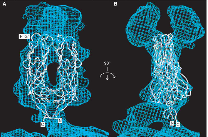
Figure 4.

The VP5* antigen domain dimer fit to the molecular envelope of the primed spike. The molecular envelope of an approximately 12 Å resolution electron cryomicroscopy image reconstruction of a VP4 spike on a trypsin-primed SA11-4F rotavirus virion is contoured at 0.5 σ. (A) Depicted from the perspective of Figures 1A and 3A. The Cα trace includes residues T259-N477 of one subunit (on the left) and residues T259-S476 of the second subunit (on the right). The termini and the F″G′ loop of one subunit are indicated. (B) Rotated 90° about a vertical axis from panel (A).
