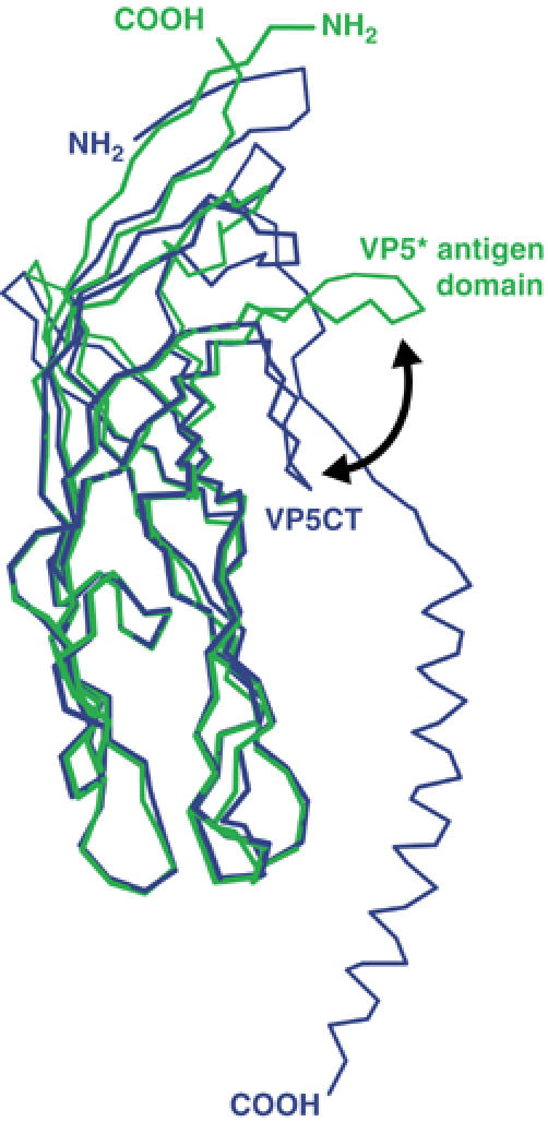
Figure 5.

Superposition of the VP5* antigen domain and VP5CT. Residues S260-S476 of a VP5* antigen domain subunit in the dimer conformation are drawn as a green Cα trace. Residues I254-L522 of a VP5CT subunit are drawn as a blue Cα trace. The black arrow indicates the movement of the GH loop between the two states.
