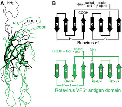
Figure 6.

Comparison of the rotavirus VP5* antigen domain and the reovirus σ1 knob. (A) Superposition of Cα traces. Residues Y298 to T455 of reovirus σ1 (black; PDB identification code 1KKE) are superimposed on residues I254-L473 of a subunit of the rotavirus VP5* antigen domain trimer (green). The residues used to calculate the superposition matrix are drawn with thick lines. (B) Folding diagram of the β-barrel of reovirus σ1 and the β-sandwich of rotavirus VP5*. Arrows depict β-strands.
