
Figure 1.
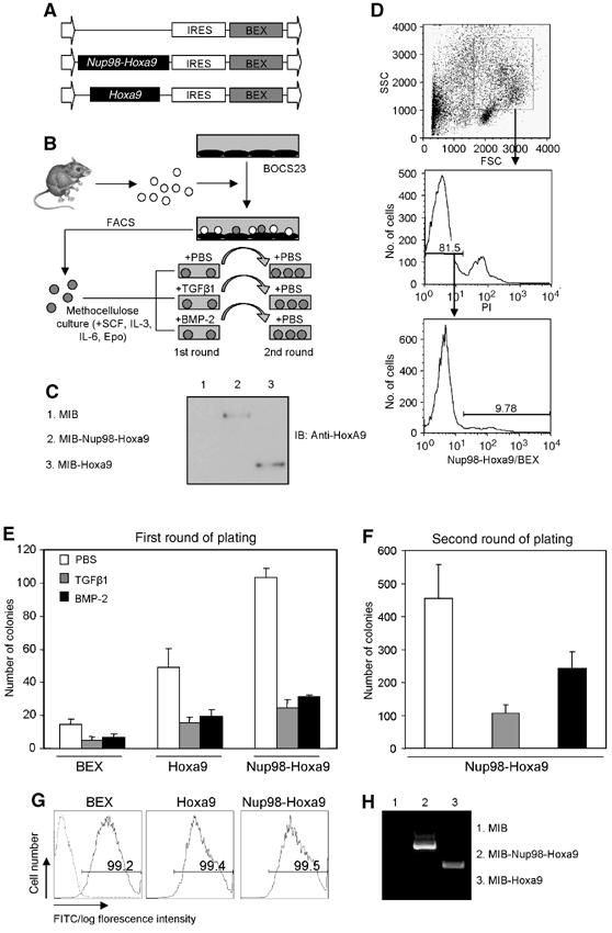
TGFβ/BMP inhibits bone marrow transformation capability of Hoxa9 or Nup98-Hoxa9. (A) Diagram of retroviral constructs expressing Hoxa9 and Nup98-Hoxa9 generated in MSCV. MSCV consists of long terminal repeat, IRES and BEX. (B) Schematic presentation of retroviral transduction procedures. Bone marrow cells were purified from 5-fluorouracil-injected C57BL/6-Ly5.2 mice and infected through cocultivation with transfected BOSC23 retroviral packaging cells for 24–48 h. BEX-positive cells were isolated by FACS and then grown in methylcellulose culture with various treatments as indicated. (C) Western blot analysis of BOSC23 cells transfected with MIB-Hoxa9 or MIB-Nup98-Hoxa9 as detected with an anti-Hoxa9 polyclonal antibody. (D) Bone marrows were gated on myeloid cells by forward scatter (FSC) and side scatter (SSC) and on propidium iodide (PI)-negative cells. Histograms indicate the percentage of BEX-positive cells that were isolated by FACS. (E) Colony numbers generated in the first plating of 2600 transduced bone marrow cells are shown. TGFβ1 (2 ng/ml) and BMP-2 (300 ng/ml) were used for treatment as indicated. Data presented are an average of at least three independent experiments with error bars. (F) Replating of 2600 transduced bone marrow cells harvested from first round of plating. Open, gray and black bars indicate treatment of PBS, TGFβ1 and BMP-2 in the first round of plating, respectively. Data presented are an average of at least three independent experiments with error bars. (G) FACS analysis of cells from second round of platings. Dash line represents nontransduced cells. (H) RT–PCR detection of the expression of the transduced genes in cells derived from the second round of platings.