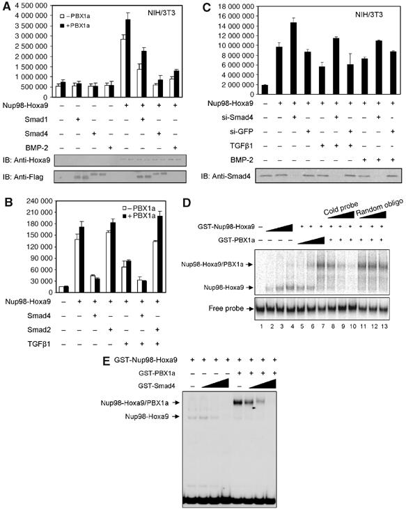
Figure 3.

TGFβ/BMP represses Nup98-Hoxa9 transcription through Smad4-mediated inhibition of Nup98-Hoxa9 binding to DNA. (A) The luciferase activities under the control of Hoxa9 promoter were measured in NIH/3T3 cells expressing Nup98-Hoxa9, in the absence or presence of PBX1a, in combination with Smad1, Smad4 or BMP-2 as indicated. Each bar represents the mean and the standard deviations of at least three independent experiments. The expression level of Nup98-Hoxa9, Smad1 and Smad4 were shown in the lower panel by Western blot. (B) Effect of TGFβ on Nup98-Hoxa9-induced transcription, in the absence or presence of PBX1a, on Hoxa9 promoter measured by Hoxa9-luc transcription reporter assay in Ba/F3 cells. Each bar represents the mean and the standard deviations of at least three independent experiments. (C) Effect of loss of Smad4 by si-RNA on Nup98-Hoxa9 transcription on Hoxa9-luc. Western blot (bottom) confirms the absence of Smad4 in si-RNA Smad4-transfected cells. (D) EMSA. Nup98-Hoxa9 alone dose-dependently binds to Hoxa9 promoter (lanes 2–4). PBX1a supershifts this protein–DNA complex (lanes 5–7). Increasing amount of specific (lanes 8–10) but not of nonspecific cold competitor (lanes 11–13) at 0, 50 × and 100 × molar excess of radiolabeled probe inhibits binding of Nup98-Hoxa9 to Hoxa9 promoter probe. (E) EMSA. Smad4 dose-dependently inhibits the DNA-binding ability of Nup98-Hoxa9 to Hoxa9 promoter probe, whether in the absence or in the presence of PBX1a, at molar ratios of 0.2, 0.4 and 1 of Smad4 compared to Nup98-Hoxa9.
