FIG. 6.
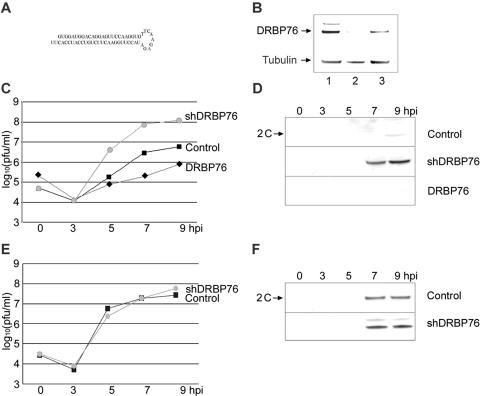
DRBP76 depletion enhances PV-RIPO propagation in HEK-293 cells. (A) Schematic depiction of shRNA targeting the DRBP76 mRNA. (B) Western blot analysis of control cells (lane 1), shDRBP76 cells (lane 2), and shDRBP76 cells transfected with DRBP76mut DNA (lane 3) using α-DRBP76 and α-tubulin antibodies as indicated. One-step growth curve analysis of PV-RIPO (C) and PV (E) propagation in control cells (▪), shDRBP76 cells (•), or shDRBP76 cells transfected with pDRBP76mut DNA (⧫) is shown. Western blot analysis of PV-RIPO (D) and PV (F) proteins in infected cell lysates at specified hours postinfection using α-2C antibody is shown.

