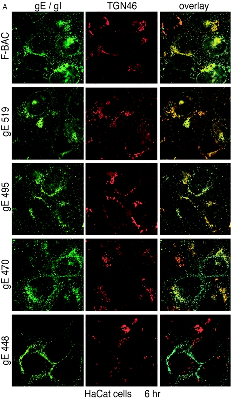
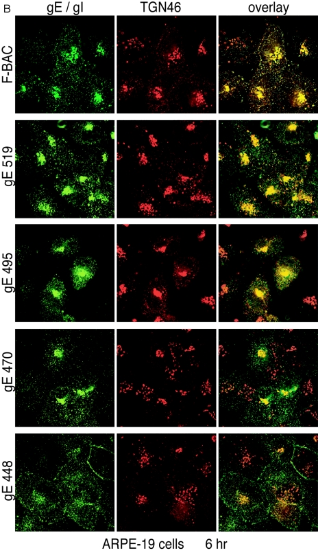
FIG.5.
Accumulation of gE/gI in the TGN. HaCaT cells (A) or ARPE-19 cells (B) were infected with F-BAC, F-BACΔgE, gE-448, gE-470, gE-495, or gE-519. After 6 h, the cells were fixed, permeabilized, and then stained with MAb 3114 (anti-gE [green]) and stained simultaneously with sheep anti-TGN46 (red). They were then washed and stained with secondary antibodies, Alexa 488-conjugated goat anti-mouse IgG and Alexa 594-conjugated donkey anti-sheep IgG, and characterized by confocal microscopy.


