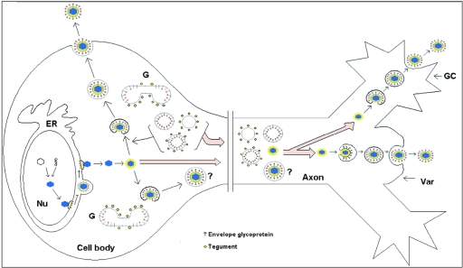
FIG. 10.
Schematic diagram of the proposed model for assembly and egress of HSV-1 in varicosities and growth cones. Unenveloped capsids coated by inner tegument proteins are transported from the cell body into axons, accumulating in varicosities (Var) or growth cones (GC). At these sites, capsids invaginate vesicles, acquiring tegument and envelope proteins. These enveloped capsids, now contained within vesicles, exit locally via exocytosis. However, the possibility that enveloped HSV-1 capsids from the cell body also enter the mid- and distal regions of the axon and exit via the varicosities and growth cones cannot be excluded. ER, endoplasmic reticulum; G, Golgi; Nu, nucleus.

