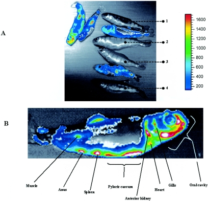
FIG. 2.
Bioluminescence imaging on live infected trout. (A) Fish were infected by bath immersion with rIHNVLUC. At 4 days postinfection, fish were subjected to a bath immersion with the EnduRen live cell substrate (Promega) and submitted to CCD imaging after being anesthetized. The luciferase activity is depicted with a pseudocolor scale, using red as the highest and blue as the lowest level of photon flux. Bioluminescence signals are displayed in pseudocolors and superimposed on the grayscale image Several controls were included under the same conditions as above: fish 1 was mock infected with no luciferase substrate bath, fish 2 was mock infected with luciferase substrate bath, fish 3 was rIHNV infected with a luciferase substrate bath, and fish 4 was rIHNVLUC infected with no luciferase substrate bath. On the right, the color code is presented. (B) A closer look at an rIHNVLUC-infected fish. Infected organs are indicated.

