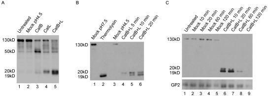
FIG. 2.
Cleavage of Ebola virus GP by CatB and CatL. (A) VSV-GP was treated with no enzyme (mock) or 20 μg/ml CatB, CatL, or CatB plus CatL (CatB+L) at pH 4.5 with 4 mM dithiothreitol for 10 min at 37°C. Reactions were quenched, and samples were analyzed by Western blotting for GP1. (B) VSV-GP was treated with 20 μg/ml CatB plus CatL in the above conditions for 5, 10, or 20 min or with 0.5 mg/ml thermolysin at pH 7.5 for 20 min. Mock treatments were performed in the same buffers containing no enzyme. Reactions were quenched and analyzed by Western blotting for GP1. (C) VSV-GP was mock treated or treated with 25 μg/ml CatB plus CatL as described above for 10, 20, 60, or 120 min. Reactions were quenched and analyzed by Western blotting for GP1 and GP2. The variable staining intensities of the 130-kDa versus the 19-kDa forms of GP1 could be due to the polyclonal GP1 antibody reacting more strongly with the 19-kDa form or to differing efficiencies of transfer between the high- and low-molecular-mass forms.

