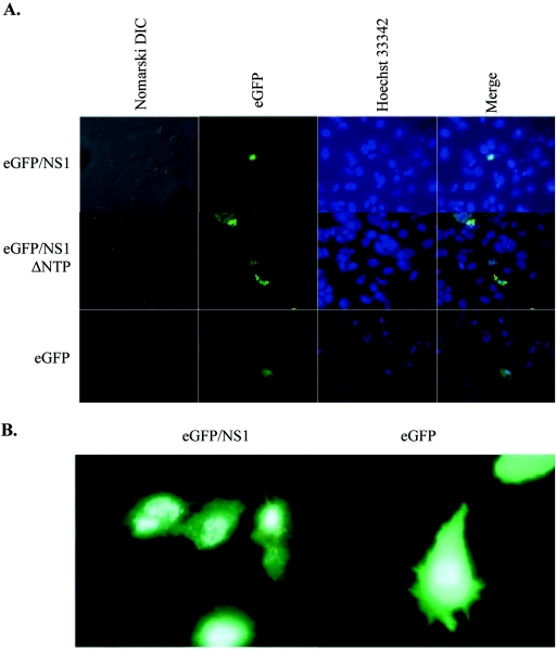
FIG. 3.
Nuclear localization of wild-type and mutant NS1. (A) eGFP/NS1-, eGFP/NS1ΔNTP-, and eGFP-transfected cells were visualized by fluorescence microscopy. The proteins are visible as green fluorescence. eGFP/NS1- and eGFP/NS1ΔNTP-transfected cells show punctuate nuclear staining, while fluorescence is distributed throughout the cell in the eGFP-transfected cells. (B) True-color image clearly showing punctuate nuclear fluorescence of eGFP/NS1 and widespread fluorescence in eGFP-expressing cells.

