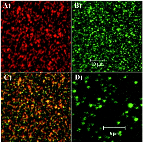
FIG. 6.
nsP3/GFP-containing structures partially purified from a nuclear pellet. The nuclear pellet after the low-speed centrifugation was further purified in a discontinuous sucrose gradient and fractionated by using the NE-PER nuclear and cytoplasmic extraction kit (Pierce) and additional centrifugation (see Materials and Methods). Fraction collected between 20 and 60% sucrose was bound to positively charged glass slides and then stained with nsP2-specific antibodies (A). Panel B shows GFP fluorescence in the sample used for panel A, panel C shows a merged image of panels A and B, and panel D is an enlarged image of that presented in panel B.

