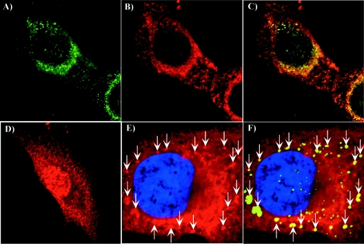
FIG. 9.
Analysis of nsP3/GFP colocalization with Hsc70 and vimentin during virus replication. BHK-21 cells were infected with SIN/389 virus at an MOI of 20 PFU/cell. At 8 h postinfection, cells were stained with antibodies specific to Hsc70 (A, B, and C) and vimentin (panels E and F) and analyzed on confocal microscope as described in Materials and Methods. Panel D indicates mock-infected BHK-21 cells that were stained with Hsc70-specific antibodies. Green staining indicates nsP3/GFP distribution and red indicates the distribution of Hsc70 (B, C, and D) and vimentin (E and F). Merged images are presented in panels C and F. Arrows indicate positions of vimentin patches colocalized with nsP3/GFP-containing structures. Nuclei in panels E and F were stained with SYTOX Orange.

