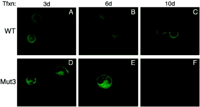
FIG. 5.
Analysis of WT agnoprotein and its phosphorylation mutant (Mut3) by immunocytochemistry. SVG-A cells, transfected with either Mad-1 WT or Mad-1 Mut3 viral DNA, were fixed with cold acetone at the indicated time points and incubated with a polyclonal anti-agnoprotein primary antibody overnight as described in Materials and Methods. Cells were then washed four times with PBS-0.01% Tween 20 for 10-min intervals and incubated with an anti-rabbit fluorescein isothiocyanate-conjugated goat secondary antibody for 45 min. Finally, the slides were washed three times with PBS, mounted, and examined with a fluorescence microscope to detect agnoprotein.

