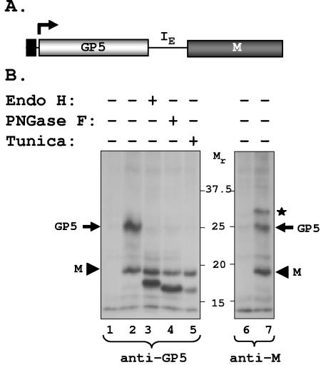
FIG. 1.
Transient expression of PRRSV GP5 and M protein. (A) Schematic of the bicistronic construct showing the GP5 and M coding regions flanking the IRES from EMCV (IE). The coding regions are under the control of the T7 RNA polymerase promoter (black rectangle) present immediately upstream of the GP5 coding region. The bent arrow shows the position and direction of transcription by T7 RNA polymerase from the vector. (B) Expression of GP5 and M proteins in cells transfected with the bicistronic vector. Mock-transfected (lane 1) or plasmid-transfected cells (lanes 2 to 7) were radiolabeled as described in Materials and Methods and immunoprecipitated with anti-GP5 antibody (lanes 1 to 5) or anti-M antibody (lanes 6 and 7). Immunoprecipitated proteins were left untreated (−) (lanes 1, 2, 6, and 7) or treated (+) with Endo H (lane 3) or PNGase F (lane 4) and analyzed by electrophoresis. Lane 5 contains immunoprecipitated proteins from transfected cells treated or not with tunicamycin. The mobility of proteins is shown in kilodaltons. The asterisk identifies a cellular protein that coimmunoprecipitates with anti-M antibody.

