FIG.1.
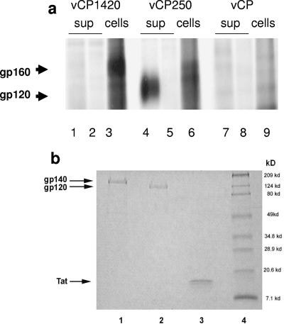
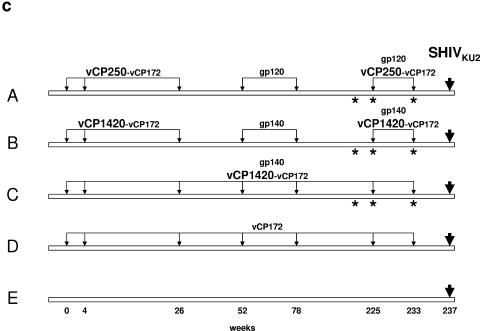
(a) Expression of HIV-1IIIB Env proteins in chicken embryo fibroblasts infected with vCP 1420 and vCP250 ALVAC-HIV-1 vaccines. Chicken embryo fibroblasts were infected with vCP1420 (lanes 1 to 3), vCP250 (lanes 4 to 6), and mock virus (lanes 7 to 9) and labeled with [35S]methionine overnight. Cells (lanes 3, 6, and 9) and supernatants (lanes 1, 2, 4, 5, 7, and 8) were lysed and immunoprecipitated with normal human serum (lanes 2, 5, and 8) or serum from an HIV-1-infected individual (lanes 1, 3, 4, 6, 7, and 9) and analyzed by SDS-PAGE, as described in Materials and Methods. (b) PAGE profiles of Env and Tat proteins. Proteins were heated at 95°C for 5 min with SDS sample buffer under reducing conditions, electrophoresed on 10 to 20% gradient gel, and stained with Coomassie blue. Lane 1, gp140; lane 2, gp120; lane 3, Tat; lane 4, molecular weight marker. (c) Schematic representation of the study design. The asterisks designate the time of Tat administration in half of the animals in groups A, B, and C.


