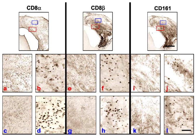
FIG. 8.

Comparison of the CD8α, CD8β, and CD161 immunoreactivities in serial sections of Lewis rat brains bearing microscopic tumors surviving treatment with RAdhsFlt3L 3 months post-CNS-1 cell implantation. CD8β-positive cells are present in greater numbers than CD161-positive cells in serial brain sections, suggesting a greater role for CD8β CTLs in inhibiting tumor growth. However, if the numbers of CD8β-positive and CD161-positive cells are combined, the total number of cells does not account for the total number of cells represented by the CD8α staining, which means there is a third cell population, most probably CD8α positive. Red letters (a, b, e, f, i, j) indicate low- (a, e, i) and subsequent high- (b, f, j) magnification images taken from the red boxed areas in each ICC staining condition shown in the top row. Blue letters (c, d, g, h, k, l) indicate low- (c, g, k) and subsequent high- (d, h, l) magnification images taken from the blue boxed areas in each ICC staining condition shown in the top row. Two areas are illustrated to highlight the heterogeneity encountered within the tumors of long-term survivors. Such heterogeneity was found only in these animals. The scale bar for the low magnification images, shown on the right-hand side panel, equals 0.5 mm; the scale bar for all other images, shown on the lower right panel, equals 50 μm.
