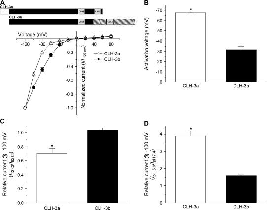
FIGURE 3.
Voltage, Cl−, and H+ sensitivity of wild-type CLH-3a and CLH-3b. (A, B) I-V relationships and activation voltages. Whole cell Cl− currents were evoked by stepping membrane voltage for 1 s between −120 mV and +80 mV in 20-mV increments from a holding potential of 0 mV. Each test pulse was followed by a 4-s interval at 0 mV. I-V plots are normalized to the steady-state current measured at −120 mV. Values are means ± SE (n = 6–7). *p < 0.0001 compared to CLH-3b. A schematic diagram of the wild-type CLH-3a and CLH-3b channels is shown above the I-V plots. (C) Effect of reducing bath Cl− concentration from 92 mM to 12 mM on whole cell current amplitude measured at −100 mV. Current amplitude was normalized to that measured at 92 mM Cl− (I92 Cl). Values are means ± SE (n = 8–9). *p < 0.0004 compared to CLH-3b. Relative current for CLH-3b is not significantly different from 1 (p > 0.2). (D) Effect of bath pH on whole cell current amplitude measured at −100 mV. Current amplitude was normalized to that measured at pH 7.4 (IpH 7.4). Values are means ± SE (n = 8–9). *p < 0.0001 compared to CLH-3b. CLH-3a requires stronger hyperpolarization for activation compared to CLH-3b. In addition, CLH-3a is more sensitive to extracellular H+ and is inhibited by reducing bath Cl− concentration.

