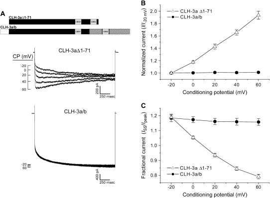
FIGURE 4.
Effect of depolarizing CPs on CLH-3aΔ1-71 and CLH-3a/b. (A) Representative current traces from HEK293 cells cotransfected with GFP and mutant channel cDNAs. Voltage clamp protocol was the same as that shown in Fig. 2 A and described in Fig. 2 legend. A schematic diagram of the CLH-3aΔ1-71 and CLH-3a/b mutant channels is shown above the current traces. (B) Effect of CP on hyperpolarization-induced current activation. Peak current amplitude was measured between 170 ms and 270 ms after stepping to −120 mV. Current values were normalized to that measured following a conditioning pulse of −20 mV (i.e., I−20 mV). Values are means ± SE (n = 8–10). (C) Effect of CP on current inactivation. Mean normalized pseudo-steady-state current (ISS) was measured over the last 20 ms of the −120 mV test pulse and normalized to peak current (Ipeak) amplitude. Values are means ± SE (n = 8–10). Predepolarization induces current potentiation and inactivation only in CLH-3aΔ1-71.

