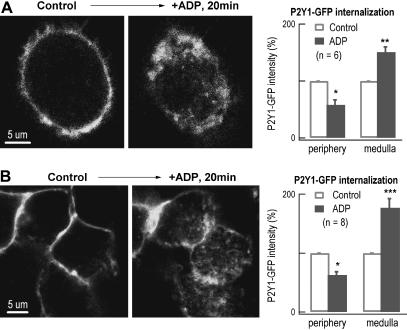
FIGURE 6.
Evidence for internalization of P2Y1 receptors by ADP-induced endocytosis. (A) P2Y1 receptor internalization by ADP stimulation in transfected DRG neurons. In an optic slice (1 μm thick) of a representative transfected DRG neuron, the P2Y1-GFP receptors were overexpressed on the cell surface (left panel). Twenty minutes after applying ADP, another optical slice was taken at the same position of the cell. The P2Y1-GFP fluorescence translocated from the periphery toward the medulla (middle panel). Statistically, compared with their initial GFP-intensities, the GFP fluorescence was decreased by 42 ± 9% on the cell surface (within 2 μm from the surface) and increased by 51 ± 9% in the medulla after ADP stimulation (n = 6, right panel). (B) P2Y1 receptor internalization by ADP stimulation in transfected HEK293 cells. In an optic slice (1 μm thick) of the HEK293 cells, the P2Y1-GFP receptors were overexpressed on the cell surface (left panel). Twenty minutes after applying ADP, another optical slice was taken at the same position of the cells. The P2Y1-GFP fluorescence translocated toward the medulla after applying ADP for 20 min (middle panel). Statistically, the GFP fluorescence was decreased by 37 ± 6% on the cell surface and increased by 77 ± 16% in the medulla after ADP stimulation (n = 8, right panel).

