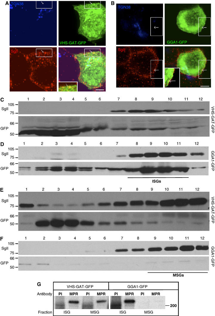
Figure 2.

VHS-GAT-GFP is found on ISGs, and inhibits removal of MPR from ISGs. PC12 cells transfected with (A) VHS-GAT-GFP (green) or (B) GGA1-GFP (green) were fixed after 24 h and labeled with anti-TGN38 (blue) and anti-SgII (red) Abs and secondary Abs. Channel merges are shown in bottom right of each panel. Insets in the merged channels are the boxed region magnified. Arrows, GFP and SgII-positive, but TGN38-negative structures. Bar=2 μm. (C–F) Fractions from EGs, which were loaded with (C, D) VG fractions 2–4, or (E, F) fractions 5–7 prepared from PNS of PC12 cells transfected with VHS-GAT-GFP (C, E) or GGA1-GFP (D, F). Proteins precipitated from the EG fractions were subjected to SDS–PAGE and immunoblotting using anti-SgII (upper panels) and anti-GFP (lower panels) Abs. Position of ISGs and MSGs on EGs is indicated. (G) ISG and MSG fractions prepared from [35S]-Met/Cys-labeled FACS-sorted cells expressing VHS-GAT-GFP or GGA1-GFP were subjected to IP with anti-MPR ab, or a preimmune control (PI).
