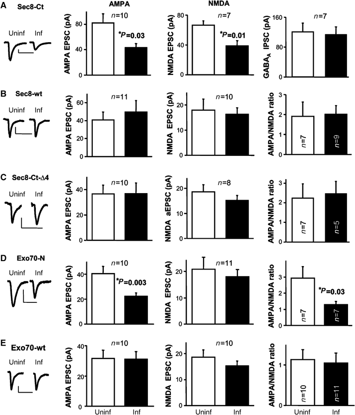
Figure 1.

Effect of different exocyst subunits on synaptic transmission. (A–E) Insets, sample trace of evoked AMPA-receptor-mediated synaptic responses recorded at −60 mV, from uninfected and infected cells. Scale bars, 20 pA and 40 ms. Left: average AMPAR-mediated current amplitude (i.e. the peak of the response recorded at −60 mV) from infected (inf) neurons expressing Sec8-Ct (A), Sec8-wt (B), Sec8-Ct-Δ4 (C), Exo70-N (D) or Exo70-wt (E), and control neighboring cells not expressing the recombinant protein (uninf). n represents the number of cell pairs. Middle: average NMDAR-mediated current amplitude (recorded at +40 mV at a latency when AMPA responses are fully decayed, 60 ms) from uninfected and infected cells (n also represents the number of cell pairs). Right: average GABAA receptor-mediated current amplitude measured at 0 mV (A) or average AMPA/NMDA ratios for uninfected and infected cells (B–E).
