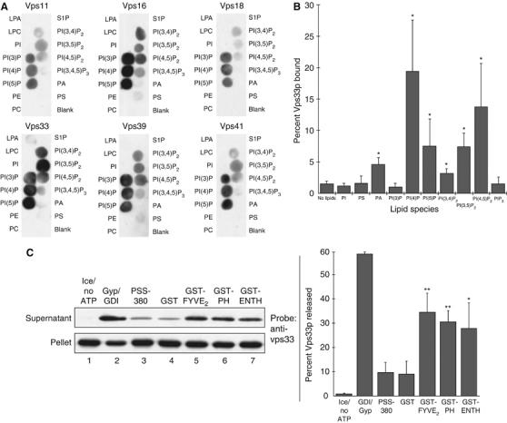
Figure 6.

The HOPS complex binds phosphoinositides. (A) PIP MicroStrips (Echelon) were blocked with HBSTG (20 mM NaHEPES, pH 7.8, 3 mg/ml defatted BSA, 200 mM NaCl, 0.1% Tween-20, 5% glycerol) and incubated with pure HOPS (0.5 ml of 0.33 nM HOPS in HBSTG) at 4°C. Strips were washed four times with HBSTG and probed with antibodies in HBSTG, then washed four times with HBSTG; secondary detection was carried out with HRP-conjugated donkey anti-rabbit antibodies (GE). Strips were washed four times with HBSTG and bound antibody was detected by enhanced chemiluminescence (GE). (B) HOPS binding to liposomes was assayed by a modification of a described method (Matsuoka et al, 1998). Liposomes with PI were made by mixing CHCl3 solutions of soy PC, soy PE, soy PI (Avanti), and rhodamine-DHPE (Invitrogen) at molar ratios of 50:25:25:0.025 and drying with N2 gas and vacuum. Lipids were resuspended in 250 μl RB (20 mM NaHEPES, pH 7.8, 100 mM NaCl, 5% glycerol, 250 mM sorbitol; 8 mM total lipids), and subjected to freeze–thaw cycles (liquid N2/bath sonication) until the suspension was clear. Liposomes with DOPA, brain PS (Avanti), or phosphoinositides (Echelon; in 1:2:0.8 CHCl3:MeOH:H2O) were prepared in the same way, with 20–25% of the PI replaced by the appropriate phospholipid. Liposomes (20 μl) or RB were mixed in 7 × 20 mm tubes (Beckman) with 10 μl pure HOPS (2.6 nM final) and 10 μl of HBSG (20 mM NaHEPES, pH 7.8, 200 mM NaCl, 5% glycerol, 250 mM sorbitol) with 1.8 M sucrose and 2 mg/ml defatted BSA, then incubated on ice for 30 min. This mixture was overlayed with 75 μl of HBSG with 0.5 M sucrose, 75 μl of HBSG with 0.3 M sucrose, and 50 μl of HBSG, then centrifuged (50 000 r.p.m., 1 h, 4°C, TLS-55 rotor). Liposomes (40 μl) were taken from the border between the top layers, and bound HOPS was quantified with a ChemiDoc system (UVP) after SDS–PAGE and immunoblotting for Vps33p, with a standard curve of pure HOPS. Liposome recovery was estimated using rhodamine-DHPE fluorescence (λex/λem 510/580 nm); reported bound HOPS was corrected for liposome recovery. Mean percentages and standard deviations for at least three independent experiments are shown. *P<0.05 for differences relative to binding to PI liposomes, by one-way ANOVA. (C) HOPS release assay: fusion assays were supplemented as indicated and incubated on ice or at 27°C for 90 min, then sedimented (16 000 g, 15 min, 4°C). Supernatants and pellets were subjected to SDS–PAGE and immunoblotting for Vps33p. Blots were quantified by densitometry. Left, representative immunoblot. Right, mean percentages and standard deviations of Vps33p released in three independent experiments. **P<0.01, *P<0.05, for differences relative to release in the presence of GST, by one-way ANOVA.
