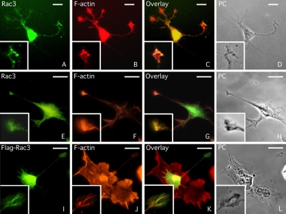
Figure 1.
Rac3 colocalizes with F-actin in the neurites and in several growth cones of neuronal cells. (A-D) Immunofluorescence staining of embryonic rat hippocampal neurons with TRITC-conjugated phalloidin to reveal the F-actin distribution and with goat anti-Rac3 antibodies followed by FITC-conjugated anti-goat antibodies. (E-H) Human neuroblastoma SK-N-BE cells were differentiated by three days of RA treatment and stained as described above. (I-L) SK-N-BE cells were transfected with FlagRac3 expression vector that stimulates the formation of long and branched neurites. NB cells were stained with TRITC-conjugated phalloidin and immunostained with FITC-conjugated anti-FLAG antibodies. The same field is shown in phase contrast (PC). In each photogram, higher magnification of a growth cone is shown. Bar, 10 μm.

