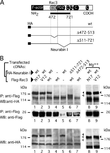
Figure 3.
The Rac3-binding region is located close to the PDZ domain of Neurabin I. (A) Schematic representation of Neurabin I protein showing the F-actin binding, the PDZ, and the coiled-coil domains. The Rac3-binding domain (Rac3 BD) is shown in gray. A solid bar indicates the 250-amino acid-long fragment of Neurabin I (a.a. 472-721) isolated by yeast two-hybrid system. The full-length wild-type and the deletion mutants of HA-tagged Neurabin I cDNA are also depicted. (B) HEK293 cells were cotransfected with the indicated HANeurabin I and FlagRac3 expression vectors. Anti-FLAG-antibodies were used to coimmunoprecipitate the FlagRac3-associated recombinant proteins. Anti-HA immunoblots (top) reveal that HANeurabin I and HANeurabin I Δ511-721 associate with FlagRac3, whereas HANeurabin I Δ472-513 does not. Wild-type HANeurabin I associates with Rac3N17 and Rac3V12 with or without addition of Mg2+. Anti-FLAG immunoblots (middle) show the recovery of Flagged-proteins. Anti-HA immunoblot (bottom) is the control to show the levels of the transfected wild-type and mutated HANeurabin I proteins. Black and empty arrowheads indicate wild type HANeurabin I and HANeurabin I mutants, respectively.

