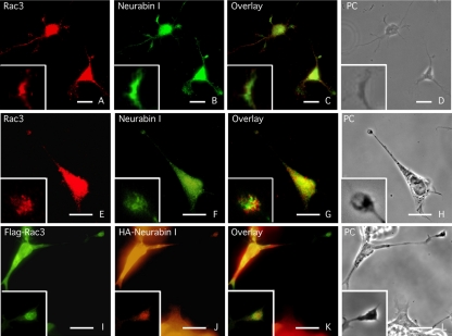
Figure 4.
Rac3 and Neurabin I colocalize in the growth cone of extending neurites. Immunofluorescence staining of Rac3 and Neurabin I proteins in primary culture of embryonic rat hippocampal neurons (A-D) and in RA-differentiated SK-N-BE cells (E-H). Cells were immunostained with goat anti-Rac3 and rabbit anti-Neurabin I antibodies. Phase contrast (PC) image of the same field is shown. Overlay and detail photograms show Rac3 and Neurabin I colocalization at the tip of neurites. (I-L) Same as above stained with FITC-anti-FLAG and TRITC-anti-HA antibodies of FlagRac3 and HANeurabin I expressing SK-N-BE cells. Bar, 10 μm.

