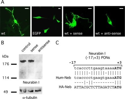
Figure 7.
Rac3-induced neuritogenesis is mediated by Neurabin I. (A) FlagRac3 expressing SK-N-BE cells were incubated alone (wt) or with sense (wt + sense) or antisense (wt + antisense) Neurabin I (-17; +3) PONs. FITC-conjugated anti-FLAG immunofluorescence reveals long and branched neurites in FlagRac3-positive cells incubated without any treatment (wt) or with sense Neurabin I (-17; +3) PONs (WT + sense). Conversely, a strong inhibition of Rac3-dependent neuritogenesis is observed upon incubation with antisense Neurabin I (-17; +3) PONs (WT + antisense). Green fluorescent protein (EGFP)-transfected cells show the morphology of undifferentiated control cells. Bar, 10 μm. (B) Anti-Neurabin I immunoblot on whole cell lysates of SK-N-BE cells incubated with sense or antisense Neurabin I (-17; +3) PONs (top). Anti α-tubulin immunoblot is to ensure equal protein loading (bottom). (C) The alignment shows the homology of Neurabin I (-17; +3) PONs between nucleotide -17 and the start codon of the Rat Neurabin I cDNA, with the human wild type (Hum-Neb) or with the HA-tagged (HA-Neb) Neurabin I.

