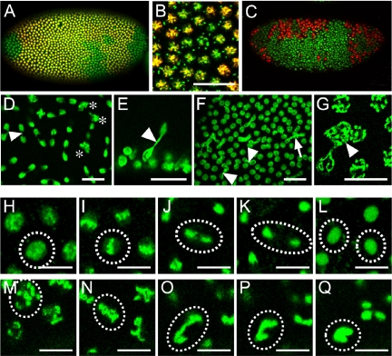
Figure 5.
Synchronicity of mitotic divisions and chromosome segregation are affected in syncytial blastoderm stage vfl mutant embryos. vfl mutant blastoderm embryos stained with anti H3-P antibodies (A-C, red) and Sytox green (A-G), vfl dsRNA injected (D-G and M-Q) and wild-type (H-L) embryos carrying H2a-GFP (H-Q). Time-lapse pictures from living embryos (see Supplemental Materials) during one round of mitosis: early prophase (H and M), metaphase (I and N), anaphase (J and O), telophase (K and P), and interphase of the daughter nuclei (L and Q). (A and B) In vfl hemizygous mutant embryos the synchronous mitotic waves are affected (A, enlarged area in B; also see Figure 2, E and F, for the wild-type pattern). (C) In some embryos, irregular areas of nuclei with highly condensed DNA are formed. (D-G) Nuclei of embryos injected with vfl dsRNA form anaphase bridges (D-F, arrowhead) or contain double sets of chromosomes (D, asterisk; G, arrowhead). Note that the two nuclei connected in E (arrowhead) originated from a perpendicular nuclear division. (H-Q) Mitotic phases of H2a-GFP-expressing nuclei (from a time-lapse movie) of a vfl dsRNA-injected embryo show strong chromatin condensation at prophase (H and M), the formation of anaphase chromatin bridges (J and O), and the failure of nuclear separation (L and Q). Bars, 25 μm in (B, D-F) and 5 μm (G-Q).

