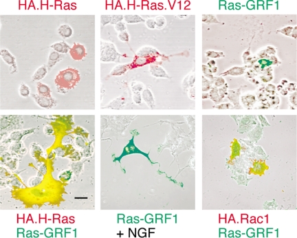
Figure 1.
Effects of Ras-GRF1 expression on the morphology of PC12 cells. PC12 cells were transfected to express the indicated constructs for 48 h. Expression of Ras-GRF1 was detected by green indirect polyclonal immunofluorescence. Expression of HA-tagged H-Ras, H-Ras.V12, or Rac1 were detected by red indirect monoclonal immunofluorescence for the HA tag. To compare morphology with untransfected cells, the fluorescence results are overlaid with a phase-contrast image of the field. Where indicated, the cultures were treated with 40 ng/ml NGF (Genentech) to induce neurite extension (bottom middle). Scale bar, 50 μm. Pictures shown are typical of results from a minimum of three independent experiments. The proportion of cells cotransfected with both H-Ras and Ras-GRF1 (bottom left) that exhibited the novel, expanded morphology was ∼25%. This morphology was never detected after transfection with either H-Ras or Ras-GRF1 alone.

