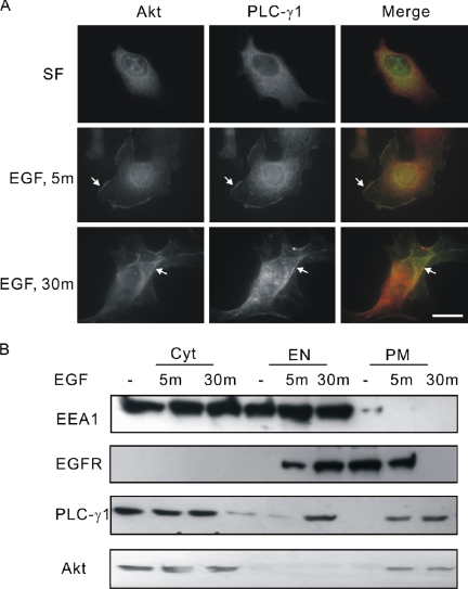
Figure 1.
Colocalization of PLC-γ1 and Akt in COS-7 cells. (A) COS-7 cells were either nontreated or treated with 100 ng/ml EGF for indicated times. The cells were then fixed and stained with anti-PLC-γ1 and anti-Akt antibodies. Colocalization (yellow) of Akt (green) and PLC-γ1 (red) is indicated by arrow. Bar, 20 μM. (B) Serum-starved COS-7 cells were either nontreated or treated with 100 ng/ml EGF for 5 or 30 min. The total cell homogenates were subcellularly fractionated into PM, EN, and Cyt. Ten percent protein of each fraction was loaded to 8% SDS-PAGE and immunoblotted with antibodies to Akt, PLC-γ1, EEA1, and EGFR.

