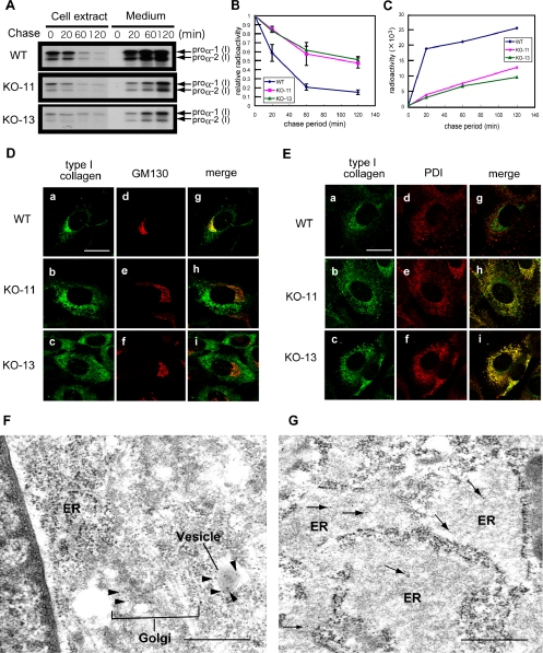
Figure 2.
Slow secretion of type I collagen and its accumulation in the ER in the absence of Hsp47. (A) Pulse-chase experiment of intracellular and extracellular type I collagen using Hsp47+/+ and Hsp47-/- cells. Immunoprecipitation was performed with specific antibody (AB765P). (B) Relative radioactivity of pulse-labeled intracellular type I collagen during chase period. (C) Radioactivity of secreted type I collagen. WT, Hsp47+/+ cells. KO-11 and KO-13, Hsp47-/- cells. (D and E) Double immunofluorescence staining was performed using antibodies against type I collagen (AB765P) and GM130 (D) or PDI (E). (a, d, and g) Hsp47+/+ cells. (b, e, and h) Hsp47-/- KO-11 cells. (c, f, and i) Hsp47-/- KO-13 cells. Bar, 20 μm. (F and G) Immunoelectron microscopy of Hsp47+/+ (F) and Hsp47-/- (G) cells using primary antibody against type I collagen (AB765P) and secondary antibody labeled with gold particles. In F, black arrowheads indicate immunogold staining of type I collagen in the Golgi apparatus and a secretory granule, and white arrows show ER. Arrows in G indicate immunogold staining of type I collagen in dilated ER. Bar, 500 nm.

