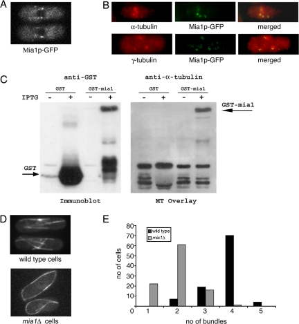
Figure 1.
Lack of the microtubule-binding protein Mia1p results in fewer microtubule bundles in interphase S. pombe cells. (A) Mia1p-GFP is enriched at the minus ends of microtubule bundles in interphase cells. (B) On MBC treatment, Mia1p-GFP localized to distinct dots around the NE coinciding with short microtubule stubs (top) and largely colocalizing with γ-tubulin-rich structures (bottom), as seen from anti-α-tubulin, anti-γ-tubulin, and anti-GFP immunofluorescence images of Mia1p-GFP cells. (C) Filter-immobilized recombinant Mia1p-GST bound taxol-stabilized microtubules in the Far Western assay. Binding was not observed with GST alone. (D) Unlike in the wild-type case, there are fewer microtubule bundles in mia1Δ cells. Shown are single maximum intensity reconstructions of α-tubulin-GFP–expressing wild-type and mia1Δ cells. (E) Histogram of the number of microtubule bundles in wild-type and mia1Δ cells (n = 100 cells).

