Figure 4.
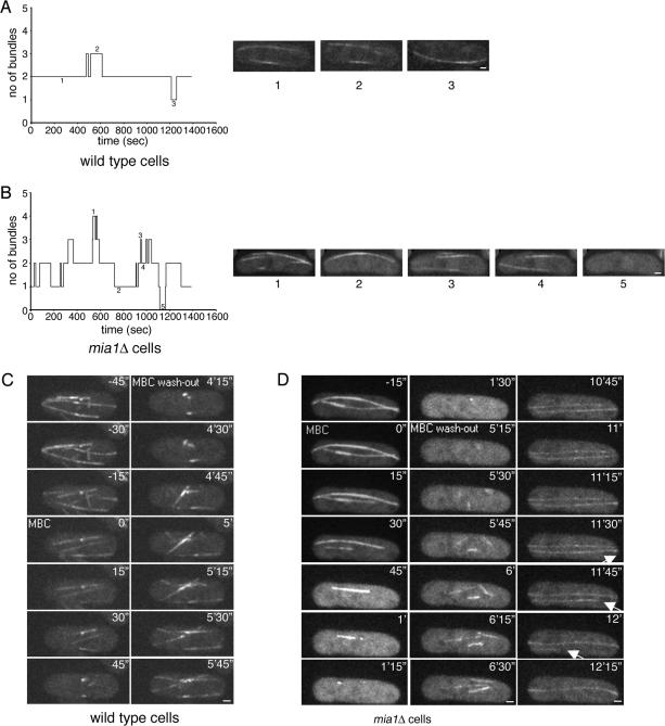
Microtubules can be nucleated from around the nuclear envelope in mia1Δ cells, but the resulting microtubule bundles are not stable. (A) Graph and several still images (1–3) following number of microtubule bundles present in one focal plane of the wild-type cell over a period of time. (B) Graph and several still images (1–5) following number of microtubule bundles present in one focal plane of the mia1Δ cell over a period of time. Data presented in A and B were obtained from time-lapse experiments using α-tubulin-GFP–expressing wild-type and mia1Δ cells (images were taken every 5 s). (C) Wild-type cells expressing α-tubulin-GFP Sid2p-GFP were mounted in the flow chamber allowing the time-lapse analysis of microtubule dynamics during MBC treatment (50 μg/ml) and subsequent washout. Microtubules depolymerized after MBC treatment leaving stable “stubs” at the NE. On washout, microtubules rapidly repolymerized from these “stubs” toward cell periphery. (D) Time lapse analysis of α-tubulin-GFP Sid2p-GFP expressing mia1Δ cell in a similar experimental setup. On washout with fresh medium, we detected efficient nucleation of microtubules from around the NE. At least one microtubule eventually detached and underwent catastrophe (as indicated by an arrow). Shown are single maximum intensity reconstructions of z-stacks. Numbers refer to the time, in minutes and seconds. Bar, 1 μm.

