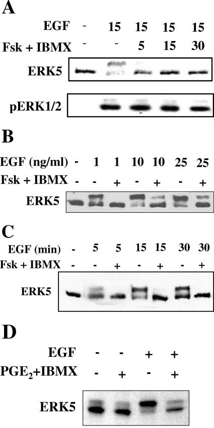
FIG. 1.
Characterization of cyclic nucleotide-dependent inhibition of ERK5. HeLa cells were grown to confluence and cultured in 0.5% FBS prior to stimulation. (A) Cells were treated with dimethyl sulfoxide (DMSO) or 10 μM forskolin and 50 μM IBMX for 5, 15, or 30 min prior to a 15-min stimulation with 10 ng/ml EGF. (Top) ERK5 activation detected by immunoblotting of a slower migrating autophosphorylated ERK5 band in cell lysates. ERK5 autophosphorylation correlates with increased ERK5 activity towards substrate (26). (Bottom) ERK1/2 activity monitored with an antibody which recognizes the dually phosphorylated active form of ERK1/2. (B) Cells were treated for 15 min with DMSO or 10 μM forskolin and 50 μM IBMX and then for 15 min with 1, 10, or 25 ng/ml EGF. ERK5 activity was monitored as described for panel A. (C) Cells were treated for 15 min with DMSO or 10 μM forskolin and 50 μM IBMX and then for 5, 15, or 30 min with 10 ng/ml EGF. ERK5 activity was monitored as described for panel A. (D) Cells were treated with 15 μM PGE2, 50 μM IBMX, and 1 mM probenecid (to reduce cAMP efflux) for 5 min. ERK5 activity was monitored as described for panel A. Results are representative of at least three independent experiments.

