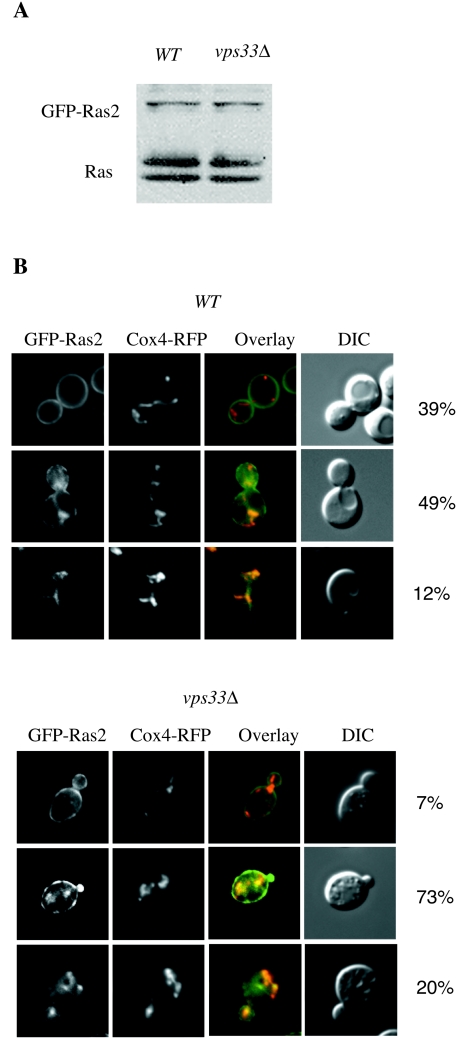
FIG. 6.
Subcellular localization of GFP-Ras2 expressed at physiological levels. (A) GFP-Ras2 was expressed from a MET25 promoter in the wild-type (LRB938) and vps33Δ mutant (RJY1654) strains. Cells were grown to exponential phase in SC-glucose medium supplemented with 12 mM methionine. The immunoblot compares the expression of GFP-Ras2 with that of the endogenous Ras1 (upper band) and Ras2 (lower band) proteins, using the anti-Ras monoclonal antibody Y13-259. (B) Wild-type (LRB938) or vps33Δ mutant (RJY1654) cells harboring MET25-GFP-Ras2 and Cox4-RFP were grown to exponential phase in SC-glucose medium supplemented with 12 mM methionine. Cox4-RFP and GFP-Ras2 were visualized by fluorescence microscopy. The percentages of cells with predominantly plasma membrane-localized fluorescence (top), plasma membrane and endomembrane fluorescence (middle), and only endosomal fluorescence (bottom) were compared. For each sample, at least six fields and approximately 150 cells were examined. A representative example is shown.

