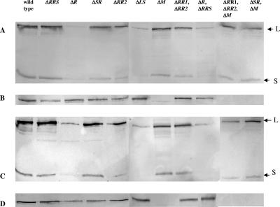
FIG. 9.
Western immunoblot analysis of form I (A and C) and form II (B and D) RubisCO accumulation in R. palustris wild-type and mutant cells grown under photolithoautotrophic growth conditions at 10 mM NaHCO3 (A and B) and 25 mM NaHCO3 (C and D). A total of 10 μg of soluble proteins was applied to each lane. Antisera raised against the form I RubisCO holoenzyme, CbbLS, reacted with a protein of approximately the same size of CbbL (form I RubisCO large subunit), giving a false-positive signal in the lane corresponding to the form I RubisCO knockout strain (ΔcbbLS); however, the absence of a signal for the form I RubisCO small subunit, CbbS, unmistakably allowed detection of the holoenzyme. The arrows point to the bands corresponding to the form I large (L) and small (S) subunits.

