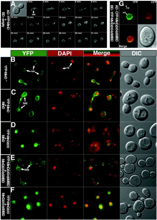
FIG. 2.
Localization of fluorescently labeled MinD proteins in yeast AH109 cells. The proteins were expressed from the relevant plasmids (Table 1) as indicated at the left of each series of micrographs (AD fusion protein in the first line and BD fusion protein, when present, in the second line). (A) Time-lapse micrographs showing a dividing nucleus at different stages of separation; time is shown in minutes at the top of each YFP image. (B through F) Columns show yellow fluorescent protein (YFP) images (green), DAPI images (red), merged images (green-red), and differential interference contrast (DIC) images (gray), as indicated. Nuclear membrane (NM) and nucleus (N) are shown by arrows. In panel B, Yfp-MinD is expressed in presence of empty-BD plasmid pGBKT7. (G) The cyan fluorescent protein (CFP) image is shown in red and the YFP image in green.

