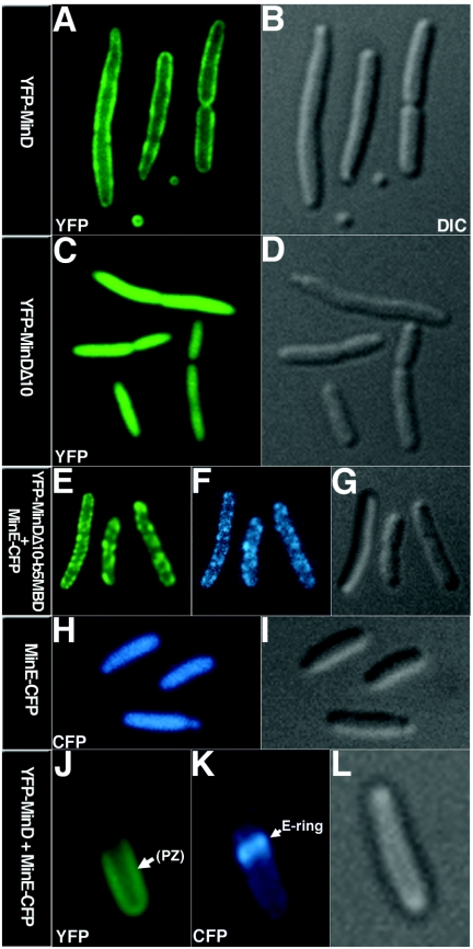
FIG. 3.
Localization of fluorescently labeled MinD and MinE in E. coli RC1. The proteins were expressed from relevant plasmids (Table 1) as indicated at the left of each image. (A, C, E, and J) Yellow fluorescent protein (YFP) images. MinD polar zone (PZ) is shown by the arrow in panel J. (F, H, and K) Cyan fluorescent protein images of MinE-CFP. MinE ring is shown by the arrow in (K). (B, D, G, I, and L) Differential interference contrast (DIC) images.

