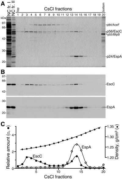
FIG. 2.
Identification of EscC and EspA as components of the EPEC NC. (A) NC fr. I (4.5 μg), NC fr. II (0.3 μg), and CsCl fractions 1 to 20 (6 μl each), shown in Fig. 1, were analyzed by 12% SDS-PAGE followed by silver staining. The positions of marker proteins are shown on the left. The positions of p56/EscC, p24/EspA, p84/AceF, and p55/BfpB are shown on the right. (B) Western blot analysis of NC fr. I, NC fr. II, and the CsCl fractions. Samples corresponding to those shown in panel A were immunoblotted with anti-EscC (upper panel) or -EspA (lower panel) antibodies. (C) Distribution of EspA and EscC in the CsCl fractions. Relative amounts of EspA and EscC in the CsCl fractions were estimated from the band densities observed on the Western blots (panel B) by using the NIH Image program. Densities (g/cm3) of the fractions were determined by refractometry.

