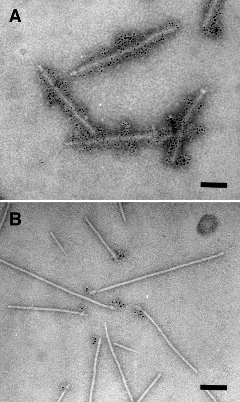
FIG. 4.
Localization of EspA and EscC in the EPEC NC. The highly purified NCs in CsCl fraction 14 (Fig. 1) were analyzed by immunoelectron microscopy. (A) EspA was detected with immunogold-labeled anti-EspA antibodies. Sheath-like structures coated with 6-nm gold particles were observed. (B) EscC was detected with immunogold-labeled anti-EscC antibodies. In contrast to EspA localization in the sheath, EscC was localized in the basal body of the EPEC NC. Bar, 100 nm.

