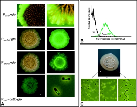
FIG. 3.
Bundles are preferential sites for sporulation. (A) Cells were spotted onto MOPS-based agar plates and incubated for 4 days at 30°C, unless specified otherwise, and fluorescence microscopic images were taken as described in Materials and Methods. The edge of a colony from strain abrB-gfp (PabrB-gfp), incubated for 14 days, was excited with blue light in a background of white light (left panel) and at a higher magnification (right panel). Strains IIA-gfp (PspoIIA-gfp) and spoIIE-gfp (PspoIIE-gfp) were captured with white light (left panel) and excited with blue light (right panel). A colony from strain cotC-gfp (PcotC-cotC-gfp) was captured by excitation with blue light (left panel). A single-cell fluorescent microscopic image of cells from strain cotC-gfp is depicted in the right panel (overlay between white light and fluorescence). Interestingly, CotC-GFP accumulates mostly at the poles of the forespore and is significantly less abundant at the flanks of the forespore. (B) Spores from strains B. subtilis 1A700 (Wt) and IIA-gfp (PspoIIA-gfp) were isolated from 5-day-old colonies grown on MOPS-based agar at 30°C. Spores were purified, and fluorescence was determined by flow cytometry as described in Materials and Methods. Green fluorescence intensity is indicated by arbitrary units (AU). (C) Single-cell microscopy analysis of a typical wild-type B. subtilis 1A700 colony grown on MOPS-based agar. Rectangles indicate the dissection sites taken for microscopy.

