FIG. 4.
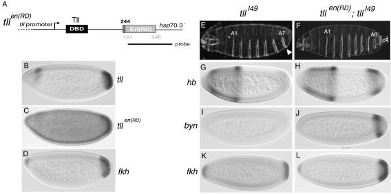
Rescue of tll mutant embryos by TllEn(RD). (A) Diagram of the tllen(RD) transgene driven by a 5.9-kb promoter region from tll. The dotted line in the promoter indicates that it is not drawn to scale. Numbers indicate amino acid positions as in Fig. 3. The probe used for the in situ hybridization shown in panel C is also depicted. DBD, DNA-binding domain. (B) Wild-type expression pattern of endogenous tll. (C) mRNA expression pattern of tllen(RD) in an otherwise-wild-type embryo. The pattern is similar to that of endogenous tll, with a visible posterior cap and a dorsal-anterior stripe. Note, however, that the transgene is also expressed at low level throughout the embryo. (D) Wild-type expression pattern of fkh. (E to L) Complementation of tlll49 mutant embryos by tllen(RD). tlll49 embryos lack structures posterior to the A7 segment (E, arrowhead) and have defective expression of hb (G), byn (I), and fkh (K) at the blastoderm stage (compare with Fig. 1C, 3I, and 3L and panel D). tlll49 embryos expressing tllen(RD) have a normal cuticle pattern (F) and restored expression of hb (H), byn (J), and fkh (L).

