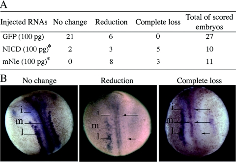
FIG. 2.
Effect of GFP, NICD, and mNle RNA injections on the production of primary neurons in Xenopus embryos. Injected embryos (stage 16) were monitored for the N-tubulin expression pattern. (A) Number of embryos that present, in the injected side compared to the noninjected side, no change, reduction, or complete loss of N-tubulin-positive neurons. *, analysis of distribution of the three N-tubulin expression profiles between NICD and GFP (χ2, 0.001 < P < 0.01) and between mNle and GFP (χ2, P < 0.001). (B) Examples of the three types of N-tubulin expression patterns obtained after RNA injections. Dorsal views are shown with the anterior end up. The injected side is shown on the right side of the images. N-tubulin expression was detected in primary neurons of medial (m), intermediate (i), and lateral (l) stripes in the noninjected side. Arrows indicate the reduction or complete loss of N-tubulin expression.

