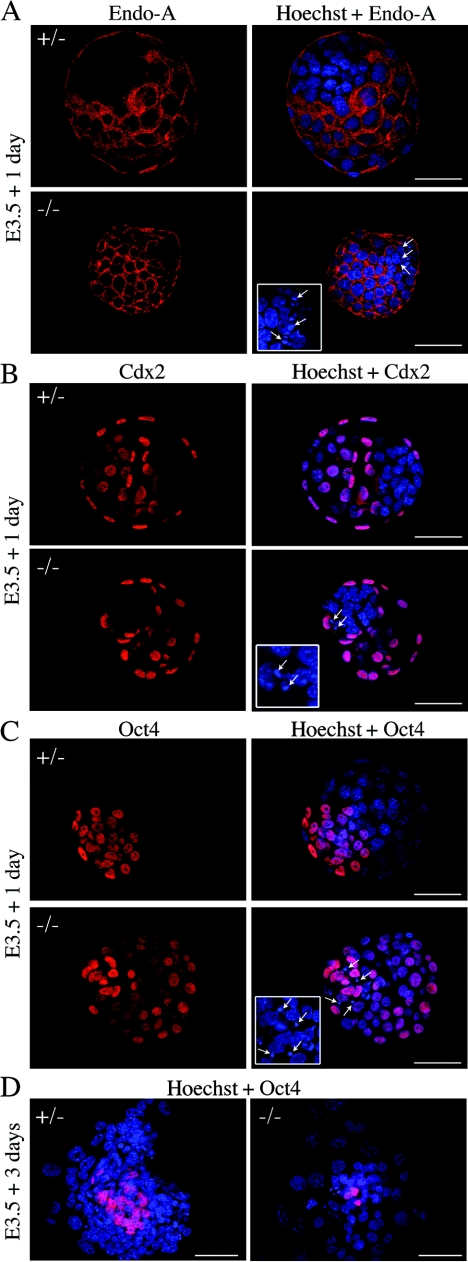
FIG. 6.
TE and ICM specification in E4.5 blastocysts and outgrowths from mNle+/− intercrosses. Immunohistochemical detection of cytoplasmic Endo-A cytokeratin (red) (A) and nuclear Cdx2 (red) (B), specific for TE, and nuclear Oct4 (red) (C), specific for ICM, in mNle+/− and mNle−/− embryos is shown. Representative control embryos (upper panel) and mNle−/− embryos (lower panel) are shown. (D) Oct4 expression in mNle+/− and mNle−/− outgrowths from blastocysts cultured for 3 days. In mNle+/− outgrowths, Oct4-positive immunostaining was detected in the ICM. In contrast, very few Oct4-positive cells were observed in mNle−/− outgrowths. Nuclei were counterstained with Hoechst stain (blue). One optical section is shown for panels A to C (flattened blastocyst morphology was due to mounting on one slide with a glass coverslip placed over it). Micronuclei were detected in the ICM of the mNle−/− blastocysts (white arrows and insets, panels A to C). Bars, 50 μm (panels A to C) and 200 μm (panel D).

