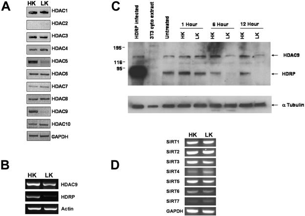
FIG. 2.
Expression of HDRP and other deacetylases during neuronal apoptosis. (A, B, and D) RT-PCR analysis was used to determine the RNA expression of indicated genes from cultures treated for 6 h with HK or LK medium. Glyceraldehyde-3-phosphate dehydrogenase (GAPDH) and β-actin were used to demonstrate that similar quantities of sample were used. (C) HDAC9 protein expression in neuronal cultures subjected to time course treatment with HK or LK medium. Lysates were obtained from cultures treated for 0 (untreated), 1, 6, and 12 h with either HK or LK medium. Western blotting was performed, and the resulting membrane was probed with an HDAC9 antibody, followed by an α-tubulin antibody to confirm equal protein loading. HDRP-infected neuronal lysates served as a positive control for HDRP expression, while 3T3 cytoplasmic extract served as a negative control.

