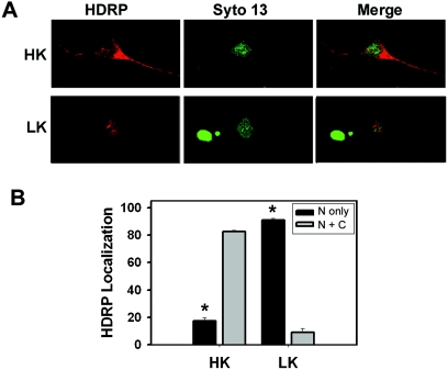
FIG. 7.
Subcellular localization of HDRP. Cultured neurons were infected with Ad-HDRP (c-Myc tag) before replacement of the medium with HK or LK medium. After 24 h of treatment, the localization of adenovirally expressed HDRP was visualized using a c-Myc antibody and a Texas Red-conjugated secondary antibody (red). Chromatin was stained with Syto 13 (green) to visualize nuclei. (A) Representative images of neurons expressing exogenous HDRP in HK and LK medium. (B) Subcellular localization of HDRP quantified in neurons treated with HK and LK medium (means ± SD; n = 3). “N only” refers to the nucleus only, while “N + C” denotes nuclear and cytoplasmic localization. *, P < 0.05 for comparisons between both HK populations and between both LK populations.

